
Mechanism of Release Of Ovum (OVULATION) with Flowchart

Mechanism of Release Of Ovum (OVULATION)

-Souti Das


  •  During the 1st 5 months of development, a finite number of primordial follicles form in the fetal ovary, these follicles contain an oocyte surrounded by follicular cells. These primordial follicles arrests at 1st meiotic division till puberty and then they further undergo development and become primary follicles.                                    

  •  During the early primary follicular stage, follicular cells are cuboidal and Zona Pellucida appears as a thin band of glycoprotein that separates oocyte and follicular cells. In the late follicular stage, follicular cells proliferate into a stratified epithelium known as Zona Granulosa.

  • During the secondary follicular stage, proteoglycan riched antrum appears in the Grannulosa layer. Cell layers in the Zona Grannulosa increase and thereby Zona Pellucida gets thickened. In this stage theca cells appear outside the follicles, those contain enzymes for androgen formation from cholesterol with the help of aromatase enzyme present in the Granulosa cells.

  • In the next stage follicular antrum increases in size, and the secondary oocyte completed its 1st meiotic division. Zona Pellucida got more prominent and various layers of corona radiate forms. With the help of LH and progesterone Graffian follicle moves toward the periphery of the ovary. New blood vessels are forms (angiogenesis) forms in the follicular cell wall, prostaglandin helps in the protrusion of blood vessels and thereby increases blood flow in the follicular cells. Leakage of plasma into the follicle causes swelling of follicular cells and after that follicular cells protruded in the ovarian capsule. At the end stigma is formed, the follicular cell is ruptured and ovum (contains Oocyte, Zona Pellucida, and Corona radiate) is released and the stigma degenerates.

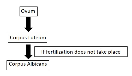
Flowchart on release of ovum


  • After release of the ovum the remaining cells of the Grannulosa and theca interna forms corpus luteum, it contains steroid producing lutin granules. Blood vessels penetrate through it and allow them to take up cholesterol, to produce progesterone. If the ovum is fertilized and implanted in the uterine wall, LH level and corpus luteum is sustained.

  • If fertilization does not occur, the cells of the corpus luteum remain active roughly for 14 days, until the level of LH fall and then involute to form corpus albicans, the secretory cells of the corpus luteum regenerate and phagocytised by macrophages and replaced by fibrous materials.


Comments

Popular posts from this blog

Are Apple Seeds Poisonous? The Truth Behind Cyanide Content

  Cyanide in Apple!! 🍏 Apple 🍎seeds are often regarded as a healthy snack that is packed with numerous nutritional benefits. However, there is a concern that apple seeds contain cyanide, a toxic substance that can cause harm to the human body. In this article, we will discuss the truth behind apple seeds containing cyanide, how much cyanide is present, and whether eating apple seeds is safe or not. The truth behind apple seeds containing cyanide Apple seeds contain a small amount of cyanide, which is a poisonous chemical compound that can be harmful to the body if ingested in large amounts. The cyanide is present in the form of amygdalin, which is a glycoside that is found in the seeds of various fruits, including apples. When apple seeds are crushed or chewed, amygdalin is released and can be converted into hydrogen cyanide, a poisonous gas. How much cyanide is present in apple seeds? The amount of cyanide present in apple seeds varies depending on the variety of apple and the ...

Principle of MTT assay

  MTT Assay Principle : The MTT assay provides a readout of cell viability & growth that works by measuring cell metabolic activity. The yellow tetrazolium MTT (3-(4, 5-dimethyl thiazolyl-2)-2, 5-diphenyltetrazolium bromide) is reduced by the metabolically active cells, by the action of dehydrogenase enzymes, to produce reducing equivalents such as NADH and NADPH. The resulting intracellular purple formazan can be solubilized and quantified by spectrophotometric means at 570nm. MTT is sensitive to light. This assay should be performed in the dark. The MTT assay measures,  cellular metabolic activity as an indicator of cell viability, proliferation and cytotoxicity.

INTRODUCTION TO RECOMBINANT DNA TECHNOLOGY(RDT) WITH ITS STEP AND APPLICATION

  INTRODUCTION TO RECOMBINANT DNA TECHNOLOGY(RDT) - Devlina Sarkar v What is Recombinant DNA technology ? RDT is a series of procedures that is used to join together (recombine) DNA segments from different organisms and inserting it into a host organism to produce new genetic combinations. v What is called Recombinant DNA ? Recombinant DNA (rDNA) is   m olecules  of  DNA  from two different species that are inserted into a host organism to produce new genetic combinations that are of value to science, medicine, agriculture, and industry. Recombinant DNA in a living organism was first achieved in 1973 by  Herbert Boyer , of the University of California at San Francisco, and  Stanley Cohen , at Stanford University , who used   E. coli  restriction enzymes to insert foreign DNA into plasmids. v What is the basic use of RDT? Using RDT we can get a huge no of copies/clones of a particular gene or DNA(cloning) by usi...